 Sometimes young parents, having come to the nextroutine examination, hear from the doctor that their baby has hip dysplasia. Many young parents are very scared when they hear about such a disease as hip dysplasia in children and begin to panic. Of course, dysplasia is a very unpleasant and serious disease, but its presence is not a reason for panic, but for treating the baby. Unfortunately, doctors often do not consider it necessary to give parents full information about this disease. And parents have a very vague idea of what they had to face. But, as you know, you need to know your enemy in person. This article will discuss what hip dysplasia is, what its consequences are and how to treat it. Hip dysplasia is usually a consequence of congenital hip dislocation. This type of congenital deformation of the musculoskeletal system is one of the most common. The damage extends to all components of the hip joint: the capsule, ligaments and muscles, the head of the femur and the acetabulum. All of these components are underdeveloped. This complication occurs in approximately 5% of all newborn babies, with unilateral hip dislocation occurring three times more often than bilateral. Girls are most susceptible to this complication – they experience congenital dislocation approximately 5 times more often than boys.
Causes of dysplasia
Doctors still haven't come to a consensus,what exactly is the cause of hip dysplasia. However, most doctors studying this problem are inclined to believe that the main cause of congenital hip dislocation is the defects of the primary embryonic organ formation, as well as the disruption of the normal process of intrauterine fetal development. There are many, many reasons for such disorders:
- Avitaminosis in a pregnant woman.
- Hormonal disorders in a future mother.
- Various infectious diseases suffered by a woman during pregnancy.
- The use of a woman during pregnancy, alcoholic beverages, narcotic substances, as well as smoking.
- Hereditary predisposition.
As studies conducted by the All-Russian Institute of Obstetrics and Gynecology have shown, children with hip dysplasia are most often born to women who suffer from:
- Early toxicosis of the first half of pregnancy.
- Late toxicosis of the second half of pregnancy - gestosis.
- Violations of water-salt metabolism in the mother.
- Violations of water-salt metabolism in the fetus.
- Disruption of the cardiovascular system.
- Rheumatic heart disease.
In 50% of all cases of birth of children withhip dysplasia during pregnancy, the babies were in a breech presentation. That is why all babies in a breech presentation undergo a thorough examination and subsequent observation by an orthopedic surgeon after birth. There is a very common misconception among young parents that sometimes congenital hip dislocations occur as a result of incorrect actions by medical personnel during childbirth. However, in fact, this is not true - in children born by cesarean section, hip dysplasia occurs no less often than in children born naturally. Congenital hip dislocation is always a consequence of the preceding subluxation of the joint. Subluxation of the joint is accompanied by hypoplasia of the acetabulum tissue, which flattens, as well as a decrease in the size of the femoral head itself, which is poorly subject to the ossification process. In this case, the femoral head often rotates its upper end forward. This phenomenon is called antetorsea. In addition, the child usually has various abnormalities in the normal development of the neuromuscular apparatus of the hip joint. Over time, the clinical picture of the disease changes: if in the first few months of the baby's life the head of the femur is displaced outward and slightly upward, then during growth there is a significant displacement upward and inward, as a result of which the joint capsule is significantly stretched and increases in size. However, in some cases of mild hip dysplasia, the displacement of the head of the femur is very insignificant. As a rule, this is observed precisely with subluxations. With true dislocations, the displacement is much more pronounced. Also, when examining a child, one can see a significant change in the structure and shape of the acetabulum and the head of the joint, as well as the joint capsule and cartilages, muscles and ligaments. Upon closer examination, one can see that the acetabulum is not just flattened, but also elongated, and its posterior upper edge is almost always practically undeveloped. As a result, the femoral head lacks the support necessary for normal functioning. The acetabulum gradually thickens the cartilaginous bottom, and excess connective tissue develops on it. If dysplasia in children under one year of age was not diagnosed in time, and treatment of the baby was not started, the course of the disease becomes much more severe with age. The child continues to develop a stable dislocation. Signs of dysplasia in children:
- The upper arch of the acetabulum often disappears almost completely.
- The hollow itself flattenes even more strongly and takes the form of a triangle.
- Due to the fact that there is no bony fence, the neck of the hip develops incorrectly - it is considerably shortened, has an obtuse cervical-diaphyseal angle.
- In addition, the neck of the hip, again because of the lack of an emphasis, turns forward.
When a baby starts walking, the load on the leg increases several times. As a result, a whole series of pathological changes occur, significantly worsening the baby's condition:
- The acetabulum continues to significantly flatten, gradually becoming almost completely flat.
- Significant changes are made and the joint capsule -one is stretched due to the forward-shifted head.
- Sometimes the articular capsule can be generally soldered to the joint bag.
- Due to the fact that the head of the hip constantly slides upwards, a slip groove is formed on the acetabulum.
- The joint cavity is in most casesdivided into three parts and takes the form of an hourglass. The upper part of the joint surrounds the head of the thigh, the middle part surrounds the cavity, but due to the fact that the depression is flattened, part of it remains hollow. The round ligament in mild cases of the disease is well expressed, but sometimes it may be completely absent.
Diagnosis of hip dysplasia
 Successful treatment of hip dysplasiajoint and the outcome of the disease largely depends on how timely the disease was diagnosed and appropriate treatment was started. If treatment is not started, complications increase like a snowball, exponentially with the time elapsed. That is why all children in the maternity hospital are examined by an orthopedic surgeon while still in the maternity hospital. As a rule, it is in the maternity hospital that congenital hip dislocation is first diagnosed. However, parents should remember that, despite the importance of early diagnosis, it is often very, very difficult to recognize it, and sometimes even impossible. Hip dysplasia in children is often well masked. That is why, even if the doctor did not find any cause for alarm, a month after birth, all babies without exception should visit an orthopedic doctor so that he can make sure that everything is okay with them. A consultation will also be necessary when the babies reach three, and then six months. Parents should never ignore these visits to the doctor - they are very important for the health of the baby - the consequences of dysplasia in children are quite severe. As mentioned earlier, all groups of dysplasias consist of several diseases:
- Congenital prelux of the hip.
- Congenital subluxation of the hip.
- Congenital dislocation of the hip.
- Radiographically immature hip joint.
In order to correctly assess the conditionhip joints of the baby, the clinical examination of the child should be carried out according to a certain method. There are a number of specific symptoms and signs by which a doctor can diagnose hip dysplasia in a child. The main ones are listed below, but parents should never try to diagnose themselves - this can lead to unpredictable complications. However, it is still necessary to know about these symptoms:
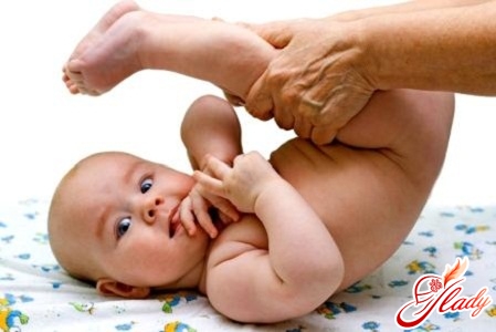
- When the legs of the crumb are diluted to the side, there is a noticeably significant restriction in the hip joints.
- Also, when the legs are bent to the side in the presence of dysplasia of the hip joint, there is a symptom of slipping or, as it is sometimes called, a click symptom.
- If the crumb is put on the tummy, you can visually visualize the asymmetry of the gluteal folds of the baby.
- With a pronounced dysplasia of the hip joints, one can see with an unaided eye the shortening of one of the legs of a crumb.
Once again it is necessary to remind that the diagnosis mustonly a surgeon-orthopedist can diagnose it. It is almost impossible to diagnose it on your own without the appropriate medical education. Also, if the baby is placed on his back, there will be a significant limitation in the hip joint when trying to spread the legs to the side. This symptom is one of the most constant and early signs of hip dysplasia. The more time passes, the older the child becomes, the stronger the limitation becomes. And if with other forms of dysplasia this symptom may not be very clearly expressed, then if the baby has a formed dislocation, this symptom will be expressed as clearly as possible. Dysplasia of the hip joints must be differentiated from other diseases in which similar symptoms are possible:
- Spastic paralysis.
- Muscular contracture.
- Congenital viral deformity of the femoral neck.
All these diseases should be completelyисключены, и только после этого врач может установить диагноз дисплазия тазобедренного сустава. Для этого ребенку необходимо провести полное обследование и внимательное изучение состояния абсолютно всех связок и мышц тазобедренных суставов, а также просто необходимо ультразвуковое исследование и рентгенограмма тазобедренных составов. Также врач при осмотре малыша обязательно учитывает влияние некоторых возрастных факторов, например, физиологическую ригидность мышц крохи. Эта физиологическая ригидность зачастую не позволяет развести ножки даже у совершенно здорового крохи. Однако подобные затруднения возникают лишь эпизодически, иногда же проблем с разведением ножек не бывает. А вот в том случае, если у малыша дисплазия тазобедренного сустава, развести ему ножки удастся только после того, как будет вправлена головка сустава. На основании совокупности всех симптомов, которые врачу удастся обнаружить при внешнем осмотре крохи, а также данных диагностического обследования малыша, и будет вынесен вердикт о наличии или отсутствии у крохи такого серьезного заболевания, как дисплазия тазобедренного сустава. Также необходимо немного подробнее рассказать про симптом соскальзывания, или щелчка. Впервые про этот симптом написал советский ортопед еще в 1934 году. Спустя некоторое время его же описал и итальянский врач, который дал симптому еще одно, третье название – симптом неустойчивости. Суть данного нарушения в том, что при попытке развести ножки крохи в сторону происходит вправление вывиха бедра. Это самое вправление вывиха и вызывает звук щелчка, который слышит врач. При подвывихах звук будет немного тише и, возможно, его не будет слышно. Однако врач обязательно почувствует его под рукой. Когда ножки малыша приводятся к средней линии, происходит повторный вывих головки, который также сопровождается звуком щелка и однократным вздрагиванием ножки. При проведении подобного исследования в обязательном порядке необходимо учитывать тот факт, что симптом щелчка чаще всего практически полностью исчезает к концу первой недели жизни крохи. Но иногда, если у малыша присутствует мышечная гипотония, он может сохраняться даже несколько месяцев, как правило, первые три – четыре. Асимметрия паховых складочек, так же, как и складок на бедрах малыша, или их различное количество на правом и левом бедре, зачастую является свидетельством того, что у крохи присутствует дисплазия тазобедренного сустава. Как правило, именно с той стороны, с которой сустав поражен, количество складок больше, и они гораздо глубже, чем со второй стороны. Расположены эти складки проксимально. Однако сам по себе этот симптом ни в коем случае не может стать основанием для диагностирования дисплазии тазобедренного сустава. Подобная асимметрия может наблюдаться даже у здоровых детишек, тогда как у одной трети детей, страдающих от дисплазии тазобедренных суставов, этого симптома не наблюдается вообще. Именно поэтому данный симптом может рассматриваться как признак дисплазии тазобедренного сустава лишь в совокупности с иными симптомами, но никак не самостоятельно. Также весьма весомым признаком дисплазии тазобедренного сустава является наличие у крохи наружной ротации крохи. Она всегда выражена достаточно сильно. Зачастую именно мамы обращают внимание на этот симптом, и именно он служит поводом для обращения за консультацией к врачу – ортопеду. Наиболее хорошо ротация заметна тогда, когда малыш спит. Как правило, ярко выраженное укорочение ножки, которое заметно на глаз, бывает в случае высокого вывиха бедра. Вопреки ошибочному мнению, ротация возможна не только при одностороннем вывихе, но и при двустороннем, в том случае, если бедра расположены на различной высоте. Некоторые мамы пытаются самостоятельно, с помощью сантиметровой ленты, определить, насколько же укорочена одна ножка. Однако у грудных детей это сделать практически нереально – у них о разнице длины ножек судят по тому, как расположены уровни коленных суставов. Для этого ножки малыша, согнутые в коленях, необходимо прижать к животику. В очередной раз обращаем ваше внимание, что все подобные манипуляции должен проводить только медицинский работник. Практически все вышеперечисленные признаки дисплазии могут быть выражены все, или же только часть, а иногда наличие их не свидетельствует о дисплазии тазобедренного сустава. Если у врача появились хоть малейшие сомнения, родители ни в коем случае не должны отказываться от более детального обследования. Если диагноз не подтвердится, и вы, и ваш лечащий врач будете точно знать, что у ребенка с суставами все в порядке. И не стоит негодовать и обвинять врача в недостаточной компетенции – это абсолютно оправданная мера предосторожности, так как не диагностированная вовремя дисплазия тазобедренного сустава может привести к тому, что у ребенка в последующем будет тяжелая инвалидность.
X-ray diagnostics
The most successful diagnosis of dysplasiahip joint in newborns using the X-ray examination method. It is performed as follows. The doctor places the baby on his back and straightens his legs. The baby's pelvis is pressed tightly against the film cassette. The baby's genitals must be protected from X-ray radiation by a specially designed lead plate. This plate, if correctly placed, does not interfere with the X-ray examination. When assessing the results of the study, the orthopedic doctor takes into account the specific age-related features of the structure of the hip joints of the newborn baby:
- In infants, the ossification of the femoral head is almost completely absent.
- The height of the femoral head of a newborn is equal to the width of the neck of the hip.
- The acetabular cavity in infants has a cartilaginous structure, so on the radiographic image it does not give a contrasting shadow.
If during normal development of the baby ossificationThe femoral head appears at about six months of age, but in children suffering from hip dysplasia, ossification does not occur before the age of one year.
Diagnosis of congenital hip dislocation in children of the senior year
 If in small children in some casesDiagnosis of congenital hip dislocation can be difficult, but from the moment the child begins to walk, it is much easier to do. In children who have reached the age of one year and are starting to take their first steps, one of the first symptoms that attract the attention of parents and doctors and allow us to assume the presence of hip dysplasia is a very late start of the walking process. Most often, this occurs in the presence of a bilateral dislocation, but sometimes it can also occur with a unilateral dislocation. Such children have a specific, characteristic gait - either the baby's instability or his lameness with a unilateral hip dislocation. If the baby has a congenital bilateral dislocation, the gait will be waddling, reminiscent of a duck. Despite the concerns of the parents, the baby does not experience any pain. The child spends as much time on his feet as healthy children, while his behavior is absolutely normal - he is cheerful, active, willingly plays. In children after one year, some symptoms of hip dysplasia also persist, but they are much less pronounced than in younger children:
- That leg, which is dislocated, as well as in crumbs, is in the position of external rotation.
- Despite the fact that there is no absolute shortening of the limb, a shortening of the limb with a dislocation is preserved.
Massage for dysplasia
If your baby has been diagnosed with dysplasiahip joint, the orthopedist will prescribe all the necessary treatment for the baby. Parents must strictly follow all the doctor's recommendations and not do anything on their own. The only thing that a mother can do, unless the doctor gives other recommendations, is a light massage. Before you start the massage, pay attention to the condition of your hands. Hands should be clean, dry and warm. Nails should be cut short, rings and bracelets should be removed - anything that can damage the integrity of the baby's skin. It is not advisable to use various massage creams intended for adults for massaging the baby - this can lead to allergic reactions in the baby in the form of skin rashes. It is much wiser to use either ordinary baby oil or baby powder. Below are described the basic massage techniques that should be done to children suffering from hip dysplasia:
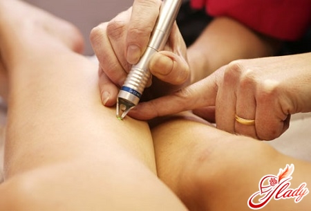
- Lay the crumb on the back, grab the whole areapalm of the thigh and begin to do light massaging movements. Make sure that your movements are not too strong. To begin massage movements it is necessary with easy stroking of a skin - watch that pressure on a skin was not considerable. The fingers should slide over the skin, but do not move it. Then the thumb and forefinger need to start making spiral movements. It is necessary to continue them for about three minutes, excluding places near the genitals. With spiral movements, pressure should be slightly increased - the skin should already be shifted. After that, place the pads of your fingers on the affected joints and massage them thoroughly, putting considerable pressure on the muscles. However, in no case do not overdo it - the crumb should not show any signs of anxiety or displeasure.
- After that gently flip the baby on the tummy and in the same way as described above, gently massage the inner thighs.
- Next, go to the gluteal area. Gently begin to rub the buttocks of the crumbs, making circular motions. After that, start not to squeeze and unclench the buttocks of the crumb slightly. During the whole time of the massage, carefully observe the reaction of the crumbs and at the first signs of discontent stop the massage.
- After you finish the massage of the buttocks,you need to go to the foot massage of the crumbs. Take the foot and begin to massage it gently from the heel to the fingers. Thoroughly massage each finger. Repeat with the second foot.
In addition to massage for hip dysplasiajoints for children, therapeutic gymnastics is extremely useful. Gymnastics, as well as baby massage for dysplasia, must be done systematically, every day. Otherwise, all your efforts will not bring any benefit to the baby's health. During the day, the mother and baby should do gymnastics at least twice a day. Each of the exercises must be repeated 10 - 15 times. Below are the basic exercises that the mother can do with the baby on her own:
- Exercise "bicycle". Put the crumb on the back, bend his legs in the lap and pull to the tummy. Act very, very carefully. Imitate the feet of a crumbs ordinary riding a bicycle.
- Exercise »bending and unbending legs. To do this, one leg of the baby in the knee, the second - completely straighten. Then change your legs. Make sure that your movements are smooth and unhurried.
- One of the legs bend in the knee and in the hipjoint, and the other press the pelvis of the crumbs and start bent legs to make circular motions. After five to six rotations, change the legs. At once you need to do two or three doses.
- Exercise "frog". Place the crumb on the back, slightly bend the legs in your lap and spread it apart. Start synchronously making circular motions with both feet.
When performing all exercises, parents mustfollow simple but very important rules. First, when spreading the child's legs to the side, be very careful and do not make sudden movements. Never spread the legs forcibly, as if overcoming an invisible obstacle. Spread the hips as much as possible without any effort. Parents should never forget about the need to visit a surgeon - orthopedist in a timely manner. The course of such a disease as hip dysplasia requires constant and careful monitoring by a doctor, who will constantly adjust the course of treatment in accordance with the peculiarity of the course of the disease in each specific case. Only constant monitoring of the course of the disease in dynamics allows you to make timely adjustments to the treatment of the child. If you have had to face such a disaster as dysplasia in infants, do not despair. Nowadays, modern medicine has learned to successfully cope even with a fairly severe form of hip dysplasia. Remember that you and your doctor have one common goal - the health of your child. Childhood dysplasia is successfully treated. That is why the first thing parents need to do is find a good orthopedic doctor whom they can trust completely. If you feel that for some reason you are unable to establish contact with your attending orthopedic doctor, try to find another specialist. Otherwise, constant conflicts with the doctor can nullify all your efforts. The health of your baby is in your hands and depends on your patience, love and perseverance! We recommend reading:
Comments
comments
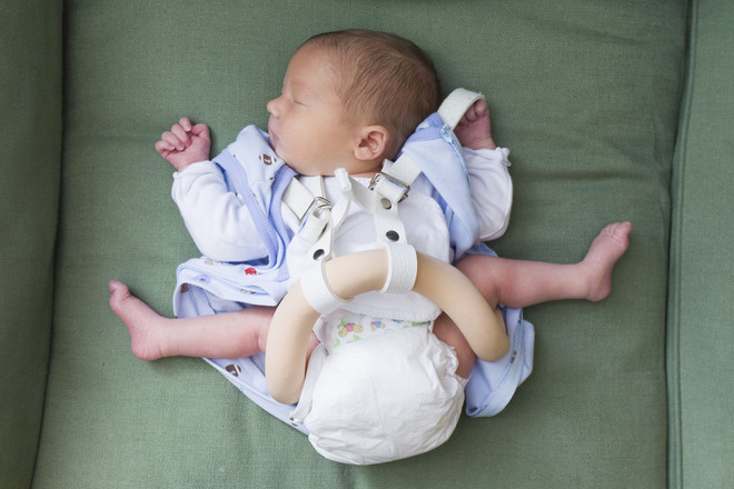 Hip dysplasia in childrenoccurs during the period of intrauterine development Photo: Getty Defects of the joint surfaces and ligaments are observed. Subsequently, this can lead to the development of hip dislocation. The pathology occurs during the period of intrauterine development of the child. Among the main reasons for its formation are:




 Sometimes young parents, having come to the nextroutine examination, hear from the doctor that their baby has hip dysplasia. Many young parents are very scared when they hear about such a disease as hip dysplasia in children and begin to panic. Of course, dysplasia is a very unpleasant and serious disease, but its presence is not a reason for panic, but for treating the baby. Unfortunately, doctors often do not consider it necessary to give parents full information about this disease. And parents have a very vague idea of what they had to face. But, as you know, you need to know your enemy in person. This article will discuss what hip dysplasia is, what its consequences are and how to treat it. Hip dysplasia is usually a consequence of congenital hip dislocation. This type of congenital deformation of the musculoskeletal system is one of the most common. The damage extends to all components of the hip joint: the capsule, ligaments and muscles, the head of the femur and the acetabulum. All of these components are underdeveloped. This complication occurs in approximately 5% of all newborn babies, with unilateral hip dislocation occurring three times more often than bilateral. Girls are most susceptible to this complication – they experience congenital dislocation approximately 5 times more often than boys.
 Successful treatment of hip dysplasiajoint and the outcome of the disease largely depends on how timely the disease was diagnosed and appropriate treatment was started. If treatment is not started, complications increase like a snowball, exponentially with the time elapsed. That is why all children in the maternity hospital are examined by an orthopedic surgeon while still in the maternity hospital. As a rule, it is in the maternity hospital that congenital hip dislocation is first diagnosed. However, parents should remember that, despite the importance of early diagnosis, it is often very, very difficult to recognize it, and sometimes even impossible. Hip dysplasia in children is often well masked. That is why, even if the doctor did not find any cause for alarm, a month after birth, all babies without exception should visit an orthopedic doctor so that he can make sure that everything is okay with them. A consultation will also be necessary when the babies reach three, and then six months. Parents should never ignore these visits to the doctor - they are very important for the health of the baby - the consequences of dysplasia in children are quite severe. As mentioned earlier, all groups of dysplasias consist of several diseases:
 If in small children in some casesDiagnosis of congenital hip dislocation can be difficult, but from the moment the child begins to walk, it is much easier to do. In children who have reached the age of one year and are starting to take their first steps, one of the first symptoms that attract the attention of parents and doctors and allow us to assume the presence of hip dysplasia is a very late start of the walking process. Most often, this occurs in the presence of a bilateral dislocation, but sometimes it can also occur with a unilateral dislocation. Such children have a specific, characteristic gait - either the baby's instability or his lameness with a unilateral hip dislocation. If the baby has a congenital bilateral dislocation, the gait will be waddling, reminiscent of a duck. Despite the concerns of the parents, the baby does not experience any pain. The child spends as much time on his feet as healthy children, while his behavior is absolutely normal - he is cheerful, active, willingly plays. In children after one year, some symptoms of hip dysplasia also persist, but they are much less pronounced than in younger children:

