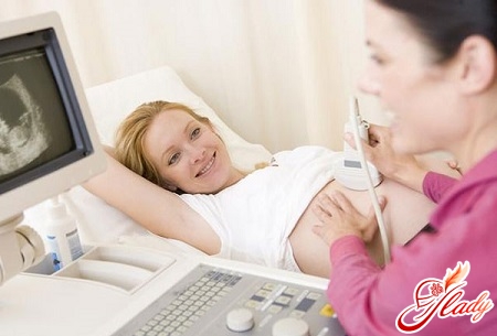
 We all dream of carrying and giving birth completelyhealthy child. But, unfortunately, an unhealthy lifestyle, poor nutrition, heredity, terrible ecology and many other factors can affect the development of the fetus. And, as a result, babies are born who sometimes do not live to be one year old. Ectopic, false pregnancy, tumors (malignant and benign) - all this poses a danger to the life of a girl. To avoid such risks, a doctor can prescribe a pregnant woman many tests, including an ultrasound. Sometimes women are afraid to undergo such tests, because they believe that this can harm the future child. In fact, there is nothing terrible about this. The main thing is not to be afraid.
What is an ultrasound study?
Ultrasound (ultrasound or echographic)(research) is often prescribed to women in the early stages of pregnancy (in this case, we are talking specifically about the first trimester). This is necessary to identify some hidden pathologies and anomalies that either pose a threat to the mother's life or affect the normal development of the fetus. The most preferable time for an ultrasound examination at this stage is from the 7th to the 14th week. It makes no sense to do it earlier, since the device may simply not see the child. By the way, abroad, doctors do not register women whose pregnancy is less than a month, since before this time the girl may have a miscarriage.
Why pregnant women need to do ultrasound
Many representatives of the fair sexI am interested in what the device can show in the first trimester, if the embryo is just beginning to develop. But it is not for nothing that doctors send expectant mothers for examination. So, let's figure out what tasks an ultrasound solves in the early stages of pregnancy, and also why you should not delay a visit to the doctor. An echographic examination helps:
- to reveal pregnancy (the fetus was fixed in a cavity of a uterus);
- to confirm that it is developing;
- to reveal ectopic pregnancy;
- To estimate, whether the size of a fetal egg corresponds to term of prospective pregnancy (gestational age is usually calculated from the first day of the last monthly);
- to determine a multiple pregnancy, if this is the case;
- To determine what wall of the uterus was attached to the chorion (subsequently, the placenta will form from it);
- to study the structure of the fetus (measuring the size of the coccyx, the thickness of the collar space to eliminate gross malformations of the fetus, etc.);
- to reveal the signs indicating the wrong development or the presence of complications in pregnancy (local increase in the tone of the uterus, the presence of a hematoma, etc.);
- The study of the yellow body, which plays a big role in maintaining pregnancy;
- to assess the condition of the appendages for the detection of the two-horned, saddle-shaped uterus, fibroids and other pathological conditions if the woman had not previously performed an ultrasound.
Conducting an ultrasound scan in early pregnancycan be performed transabdominally (the fetus is viewed through the abdomen with a full bladder) or transvaginally (through the vagina) with a sensor. In the latter case, the accuracy of the study increases. In addition, transabdominal ultrasound is contraindicated in case of urinary incontinence, diseases of the genitourinary system (cystitis, urethritis, pyelonephritis in the acute stage). Plus, it contributes to the introduction of microorganisms into the overlying sections. If the ultrasound beam is dosed accurately (taking into account the gestational age), then this type of examination does not pose a particular danger to either the mother or the fetus. Therefore, you should not be afraid of ultrasound, follow prejudices, postponing the visit for later. By the way, you will have to resort to this procedure two more times - in the second and third trimesters.
Decipher the tests correctly
Determine if there is an embryo (so calledмалыш до 8-й недели своего развития) в полости матки, на хорошем ультразвуковом аппарате возможно уже с 5-6-й недели. Параллельно врач оценивает его сердечную деятельность. Для этого он производит регистрацию частоты сердечных сокращений. В норме они колеблются от 110 до 130 уд/мин. Если же удары происходят гораздо реже, то есть становятся менее 100 ударов в минуту, необходимо срочно обратиться к гинекологу. Так как это может указывать на возможное прерывание беременности. Также неблагоприятным признаком является нахождение плодного яйца в нижней части матки. Это свидетельствует о том, что в любой момент может произойти выкидыш. Требуется срочное лечение, если у плода сердце все-таки бьется – тогда ребенка можно будет спасти. Если стук отсутствует, то это говорит о неразвивающейся беременности, которую уже не сохраняют. Здесь только один выход – искусственное прерывание. Чтобы обнаружить на ранних сроках пороки в развитии, не совместимые с жизнью, необходимо внимательно исследовать анатомию плода. До 11 недель может сохраняться так называемая физиологическая грыжа передней брюшной стенки, при которой кишечник оказывается снаружи. До 11 недель это нормально и расценивается как обычный этап развития органов и систем плода. Поэтому в данном случае паниковать не надо. Однако если такая аномалия сохраняется после этого срока, то здесь требуется динамическое наблюдение. С 11-й по 13-ю неделю важно оценивать величину воротниковой зоны. Если же она более 3 мм, вам понадобится провести определенные исследования во втором триместре, чтобы полностью исключить пороки развития плода. Такие симптомы свойственны, например, синдрому и болезни Дауна. Во втором триместре проводят УЗИ и пренатальный скриниг, для которого берется кровь из вены и исследуются определенные показатели. Очень важно в процессе ультразвукового исследования на ранних сроках беременности оценить желточный мешок, так как он обеспечивает нормальное развитие малыша. Желточный мешок представляет собой тонкостенное образование округлой формы, которое находится внутри с 6-й до 12-й недели. Плохо, если после 8-й недели его размер менее 2 мм или более 5,5 мм, а также когда он исчезает раньше 12 недели или сохраняется позже. В данном случае подобная аномалия расценивается как неблагоприятный признак. Скорее всего, это указывает на неразвивающуюся беременность и требуется проведение своевременного лечения. Повышение тонуса матки и наличие гематомы (скопления крови между плацентой и стенкой матки) – все это расценивается как признак угрозы выкидыша. В данном случае требуется срочная медицинская помощь. Если же вовремя не обратиться в больницу, то и мать, и ребенок могут погибнуть. Таким образом, УЗИ на ранних сроках беременности дает очень важную информацию, которая позволяет своевременно выявить возможные осложнения и предупредить их развитие. Что, в свою очередь, обеспечит в дальнейшем рождение здорового ребеночка и подарит вам счастье материнства. Обратите внимание: в наши дни на эхографическое исследование направляют всех беременных представительниц прекрасного пола. Поэтому не стоит переживать и паниковать раньше получения анализов. Ведь в этот период очень важно сохранять душевное и физическое равновесие. И тогда у вас в жизни будет все хорошо.


