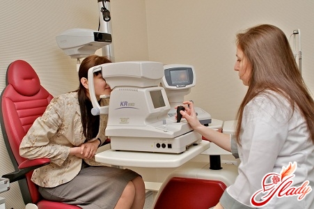
 Modern humanity suffers from the mostvarious diseases. This is explained by various factors - conditions and lifestyle, its high pace and the impact of unfavorable external factors. The human visual system is no exception to the rule. There are a huge number of different diseases of the visual system. And retinal dystrophy is far from the last place in this list. The most susceptible to this disease are elderly people, especially those suffering from such diseases as hypertension and diabetes. Abuse of alcoholic beverages and smoking also greatly increase the risk of developing retinal atrophy.
Structure of the retina
Before talking about a disease likeretinal dystrophy, it is necessary to tell our readers what the retina is and what it is for. A thin layer of nerve cells that forms the inner shell of the eye is the retina. The retina consists of two parts - the back and the front. Nerve cells - photoreceptors, designed to absorb light, form the back of the retina. From these photoreceptors, nerve impulses carrying certain information transmit it to the central nervous system, and from there - to the brain. And already there, in the visual analyzer, the actual processing of the received information occurs. In the front part of the retina, there are no such photoreceptors. Despite the fact that the retina itself is very, very thin, it consists of several layers, each of which performs its own function. For example, the outer layer of the retina, the so-called pigment layer, is very closely connected with the middle, vascular, shell of the eye. In the very center of the retina is a blind spot - the optic disc, which does not contain photoreceptors. In close proximity to the central nerve disk is the macula - a yellow spot, in the center of which is a kind of pit, in which the image is of the highest quality. That is why if there is a destructuring (change) of the retina in this place, all vision is significantly reduced. If other areas of the peripheral retina change, then the patient's field of vision is greatly narrowed, depending on the localization of the affected source.
The process of retinal dystrophy
In the event that in the cells of the retinathe normal metabolic process is disrupted, as a result of which qualitatively and quantitatively altered metabolic products accumulate in the cells, damage to the retina may occur, expressed to a greater or lesser extent - dystrophy. There are many reasons for the development of retinal dystrophy. As mentioned above, such reasons include smoking, alcohol abuse, and diabetes. Also, the reasons that can provoke the development of retinal atrophy include:
- Diseases of the circulatory system. Violation of both the composition of the blood and the circulatory process equally often can provoke the disease.
- Violation of the process of lymph circulation, including lymphadenitis, especially chronic and recurrent.
- Infectious diseases in the elderly.
- Violations of the normal hormonal background of the human body, especially caused by diseases of the endocrine system.
- Violation of the normal metabolic process - there is a disruption of nutrition of all cells, including the eye tissues, which ultimately can lead to retinal atrophy.
Strictly speaking, it is most often the violationblood circulation in the tissue cells and retinal dystrophy develops. This process is very typical for elderly people. However, in some cases, dystrophy can develop in young people. For example, this may well happen if a person suffers from severe myopia. Severe myopia leads to the fact that the transverse size of the eyes increases significantly, and accordingly, the pressure on the eye shell becomes many times stronger. In addition, eye trauma can lead to the development of retinal dystrophy, especially if the mucous membrane is damaged. Also, retinal dystrophy can be provoked by any chronic eye or cardiovascular diseases, kidney diseases, leading to frequent increases in blood pressure. And another factor provoking retinal dystrophy can be the strong impact on the human body of various toxins - simply put, poisoning.
Types of dystrophy of the retina, its signs
Doctors divide atrophic retina into twosubspecies, each of which has its own causes, features of the course of the disease and prognosis. So, dystrophy is: Central retinal dystrophy Central or, as it is also called, generalized retina occurs in the macula area - the place where vision is most distinct. The cause of the development of such a complication is almost always a pathological change in the vessels of the eyeball, most often having an exclusively age-related nature. In this case, the development of the disease is extremely slow - over several years. The clarity of vision does not disappear instantly, but gradually. A person sees especially poorly those objects that are located close to him. As a rule, if central dystrophy is not accompanied by peripheral dystrophy, complete blindness does not occur. Visible signs that a person suffers from retinal dystrophy are the presence of such signs as curvature of the image, the appearance of various spots before the eyes, most often colored. Also, these signs include double vision of objects, which such sick people complain about quite often. However, of course, we should never forget that all people are unique - some may have all of the above-mentioned features, while others may have only one of them. The central retina also has two subtypes:
- Dry form of retinal dystrophy.In this case, between the choroid and the retina of the eye, there is an accumulation of products released by the body as a result of metabolism. These secretions are a kind of tubercle on the surface of the retina. This subtype of dystrophy is the most rare - vision loss occurs extremely slowly, and the prognosis for this type of disease is the most favorable.
- Wet form of retinal dystrophy.Wet retinal dystrophy is a much more severe form of the disease – vision can be significantly reduced in a relatively short period of time – from several days to several weeks. With the wet form of retinal dystrophy, either blood or lymphatic fluid accumulates in the area.
Peripheral retinal dystrophyPeripheral retinal dystrophy affects any other areas of the retina. The particular insidiousness of this disease is that in most cases it is practically asymptomatic - only sometimes a sick person may experience flashes of bright spots before the eyes. However, most often this type of retinal dystrophy is detected completely by chance - during a routine examination by an ophthalmologist.
Treatment of retinal dystrophy
Regardless of what the reasons werecaused the development of retinal atrophy, the underlying disease requires immediate treatment. Moreover, the sooner the treatment is started, the more effective it will be. As a rule, it is not always possible to completely restore the level of vision with the help of treatment - but the sooner it is started, the higher the doctors' chances of success. First of all, to treat retinal dystrophy, it is necessary to eliminate or minimize the manifestation of those diseases that, in fact, are the cause of this disease. And only after this is done, the ophthalmologist will begin direct treatment of retinal dystrophy. Today, an aragon laser is used to treat retinal atrophy. It is used to strengthen the retina, and if the disease is already in an extremely advanced state, the laser allows you to limit retinal detachment. The principle of laser treatment is as follows - the effect of the laser entails an extremely sharp increase in temperature. The increase in temperature, in turn, leads to the fact that the tissues coagulate. The operation is absolutely bloodless. In addition, the laser has extremely high precision and allows for effective fusion of the vascular and retinal membranes of the eye. This operation is performed as follows: a special contact lens with an anti-reflective coating is placed on the affected eye of the patient. Thanks to this contact lens, the doctor is able to penetrate the eye. The laser beam itself is supplied through a special slit lamp. And the surgeon, using a stereo microscope, has the ability to focus the laser beam on the required area and control the overall course of the operation. In conclusion of the conversation about retinal dystrophy, it is necessary to remind once again that the earlier the disease is detected, the more effective its treatment. Therefore, any person should regularly visit an ophthalmologist for preventive examinations. We recommend reading:


