 Our spine resembles a pearl necklace –The vertebrae, like pearls, are connected to each other by rigid ligaments. Between the vertebrae are cartilaginous intervertebral discs, which prevent the vertebrae from touching and act as shock absorbers between them. The spine usually consists of 32-34 vertebrae, which perform different tasks and belong to different sections of the spine. In total, there are five sections in the spine:
- neck section, which consists of seven vertebrae;
- thoracic region, which consists of twelve vertebrae;
- lumbar region, which consists of five vertebrae;
- Sacrum, which consists of five vertebrae;
- The coccygeal department, which consists of three to five vertebrae.
Inside the spinal column is the vertebralcanal – a cavity formed by the arches of the vertebrae. Nerve roots, blood vessels and the spinal cord pass through the spinal canal. The human spine is adapted to walking upright, but it is walking upright that is the factor that has a detrimental effect on our spine. Intervertebral cartilages experience enormous stress every day from human movements and vibrations that occur during movement. Over time, the cartilage becomes deformed and ceases to perform its functions in full. A person begins to experience tension and back pain – characteristic signs of osteochondrosis.
Diseases of the spine
A huge number of people experience back painpeople, regardless of age. More than 80% of people at least once in their lives have faced the problem of back pain. Already at the age of 40-45, spinal diseases become one of the most common causes of loss of ability to work. The cause of various spinal diseases is a violation of the anatomical form and functional state of the spinal column. And such violations are due to the lifestyle of a modern person. Using the achievements of civilization, humanity leads a sedentary lifestyle. Most people do not need to make muscular efforts, many have an unbalanced diet, almost everyone is prone to bad habits. All this leads to degenerative and dystrophic changes in the vertebrae and intervertebral discs. Depending on what changes have occurred, this or that disease occurs. Basically, all spinal diseases have similar symptoms - pain and muscle tension, only the localization of pain differs. But osteochondrosis is the most common disease - in 90% of cases it causes back pain.
Clinical picture of osteochondrosis
Osteochondrosis - a disease causedизменением межпозвоночного хряща (chondron – означает хрящ) с сопутствующей реакцией тела позвонка (osteon – кость). Деформируясь, межпозвоночный диск уплотняется и становится тоньше. При этом происходит сжатие костной структуры тел позвонков, позвонки начинают испытывать перегрузку. Придавленные межпозвонковые диски деформируются ещё больше, в некоторых местах они начинают выпирать за границы позвоночника. Рано или поздно диск пережимает нервные корешки, что вызывает их воспаление. Так возникает болевой синдром. В зависимости от того, какой из отделов позвоночника повреждён, бывает несколько различных видов остеохондроза. Различают остеохондроз шейный, грудной, поясничный, крестцовый, распространённый (когда поражение охватывает все отделы позвоночника). Наиболее распространёнными являются поясничный (более 50% заболеваний) и шейный (более 25%) остеохондрозы. Часто встречаются случаи, когда поражаются несколько отделов позвоночника – шейно-грудной остеохондроз, пояснично-крестцовый. Начальные признаки остеохондроза позвоночника проявляются возникновением тупых болей и ломоты в пояснице (при поясничном остеохондрозе), дискомфортным напряжением мышц шеи, хрустом в шейных позвонках (при шейном остеохондрозе). Нередко возникающие при грудном остеохондрозе боли воспринимаются больными, как боли в сердце. В дальнейшем боли нередко начинают отдавать в ноги или руки; конечности немеют и становятся холодными. Нередко боль появляется даже в пальцах рук или ног. Боль в спине усиливается при резких движениях или тряске (например, во время поездки в транспорте). Становится невозможным выполнять какие-либо работы с наклоном туловища вперёд – при согнутой спине боль резко усиливается, но перейти в вертикальное положение больному удаётся не всегда. Чем больше развивается остеохондроз, тем больше ограничивается подвижность и гибкость позвоночника. Истончённые межпозвоночные диски уменьшают расстояние между позвонками, и у последних остаётся меньше пространства для движения. Кроме того, мышцы вокруг поражённого участка позвоночника постоянно находятся в напряжённом состоянии – организм пытается блокировать повреждённые позвонки для предотвращения их дальнейшей деформации. «Зажатые мышцы» доставляют дополнительные дискомфорт и боль, и способствуют ещё большему ограничению подвижности. Все перечисленные симптомы могут возникать как в состоянии покоя, так и при движении или физическом усилии (возникает дополнительное давление на нервные корешки). Диагностика остеохондроза Как же идентифицируется остеохондроз, если его симптомы на начальном этапе можно принять за симптомы других заболеваний? Конечно, врача будет интересовать анамнез. Выслушав больного и проведя осмотр, доктор направит его на дополнительное обследование. Существует несколько различных методов обследования для диагностики остеохондроза. Рентгенография Обследование позвоночника с помощью рентгена (спондилография) позволяет объективно оценить его состояние. Рентгенологические признаки остеохондроза выявляются уже на начальных этапах заболевания. Спондилография даёт представление о состоянии позвонков и косвенно – о состоянии костных каналов и межпозвонковых дисков. Снимки выполняются в прямой и боковой проекции. Если врач посчитает нужным, назначаются функциональные снимки в различных положениях – в положении боковых наклонов, в положении сгибания и разгибания. В случае необходимости больному делают томограмму – послойное рентгеновское обследование. Кроме обычного рентгенологического обследования, по особым показаниям применяют контрастные рентгеновские обследования позвоночника. К таким обследованиям относятся:
- Pneumomielography - using as a contrast 20 to 40 milliliters of air. Air is introduced into the vertebral canal after spinal puncture;
- Angiography - when 10-15 milliliters of contrast is injected into a vertebral or carotid artery, and then a series of images is taken in two projections;
- Myelography - a coloring substance is used,Introduced into the spine to isolate the structure of the ridge. With the help of myelography, you can determine the pressure of the intervertebral disc on the spinal cord. The procedure lasts for half an hour, it is produced under local anesthesia. First, the anus is lowered with an anesthetic. Then, using a thin needle into the liquid filling the space near the spinal cord, a coloring opaque substance is injected. After the contrast is injected, the X-ray table slowly tilts, and the substance moves along the spine from the lower part to the upper one. After the end of the procedure, the patient needs to spend several hours lying down.
- Discography - is similar to myelography, with the difference that the coloring substance is injected into the painful disc to determine if it is the cause of osteochondrosis.
Other methods of examination of the spineX-ray does not give the doctor a complete picture for establishing an accurate diagnosis. With its help, it is possible to reliably judge mainly the degree of wear of the vertebrae and their displacement. Unlike X-ray, a CT scan gives a clear picture by which one can judge the presence and location of an intervertebral hernia. This examination method allows you to get a clear and detailed image of the spinal column and shows all changes in it from different positions and angles. At the same time, a CT scan is a more gentle method that is easily tolerated by patients. Magnetic resonance imaging (MRI) - this method gives the most accurate image of the spine to date. This is possible due to the fact that the examination is performed not with X-rays, but with the help of a strong magnetic field. MRI is the preferred examination method because it allows you to assess the condition of the spinal canal, nerve fibers, bones, muscles, ligaments; with its help you can see any changes that occur with osteochondrosis.
Symptoms of osteochondrosis
Localization and nature of pain sensationsosteochondrosis depends on which part of the spine is affected by the disease. Of course, the signs of cervical osteochondrosis are very different from the signs of damage, for example, to the lumbar spine. And yet, there are general symptoms of osteochondrosis that will tell you that you are sick:
- making a sharp movement, turning his head tofailure, twisting the torso in conjunction with the tilt, or straightening up after the tilt - you suddenly feel a sharp and severe pain in the back, which is similar to a current stroke;
- after the "shock" you for a while seem paralyzed and freeze, unable to move;
- The muscles in the place where the pain arose are painfully strained;
- if you press your fingers in the place of the spine where you felt pain, then the feeling of sharp pain will repeat;
- the mobility of the spine becomes noticeably limited. It is difficult for you to find a position in which pain could have subsided;
- if the posture you have accepted is unsuccessful, the pain is sharply increased.
There are also symptoms that are characteristic of a certain type of osteochondrosis.
Cervical osteochondrosis
Signs of cervical osteochondrosis are easyconfused with symptoms of other diseases. When the cervical spine is affected, the pain is transmitted to the arms, the back of the head; severe headaches appear, turning into migraines. Severe, boring pain in the neck or back of the head may occur, which intensifies when turning the head, coughing, sneezing. Pain from the neck can radiate to the shoulder and side of the chest. In some cases, the patient experiences not only a headache, but also dizziness, tinnitus, and visual disturbances. If the disease progresses, persistent circulatory disorders of the brain or spinal cord are possible. When the nerve roots are compressed (squeezed) in the lower segments of the cervical spine, symptoms similar to those of angina pectoris occur - pain in the heart, neck, and shoulder blades. The pain intensifies with movement and is not relieved by cardiac drugs. The causes of cervical osteochondrosis are due to the anatomical features of this segment of the spine. The cervical vertebrae are under constant strain, holding and often turning the head, while the size of the cervical vertebrae is significantly smaller than the vertebrae of the rest of the spine. We must not forget about the narrowness of the internal spinal canal. A huge number of nerves and vessels are concentrated in the neck area, including a large vertebral artery passing inside the spinal canal, feeding the brain. All this fits tightly together in the cramped space of the cervical vertebrae. With cervical osteochondrosis with displacement of the vertebrae, the nerve root is pinched, its edema and inflammation quickly develop.
Osteochondrosis of thoracic and lumbar spine
The spine in the thoracic region together with the ribsserves as a framework that protects vital organs. The thoracic vertebrae have such a structure that they remain slightly mobile, so they are rarely subject to degradation and deformation. As a result, pain in the thoracic spine is also rare. Signs of thoracic osteochondrosis are often mistaken for manifestations of other diseases - it is confused with angina pectoris and even mistaken for myocardial infarction. When the thoracic spine is affected, the pain is girdle-like in nature, and the patient may think that it comes from the lungs, heart or even stomach. It is precisely because the signs of thoracic osteochondrosis are “masked” as other diseases that differential diagnostics are of great importance in making a diagnosis. Lumbar osteochondrosis is nothing more than changes in the intervertebral discs, located, accordingly, in the lumbar region, which consists of 5 large vertebrae. The lumbar region connects the sacrum and the thoracic region. Osteochondrosis of the lumbar spine is much more common than other types of osteochondrosis. This is explained by the fact that it is the lumbar spine that bears the entire load of the human body weight, as well as the load that a modern person has to carry every day - briefcases, shopping bags, etc. That is why patients so often go to the doctor not only with osteochondrosis itself, but also with the complications that it entails, in particular, with intervertebral hernias. Intervertebral hernias are not such a harmless phenomenon; in especially severe cases, even paralysis of the limbs is possible.
Symptoms of lumbar osteochondrosis
People who have been diagnosed by a doctor with lumbar osteochondrosis report the following complaints and symptoms:
- Painful sensations in the lumbar region, andThe pain sometimes has a shooting character and gives to the buttocks and legs. Painful sensations at slopes or squats of the sick person amplify in times. The same happens with prolonged stay in an uncomfortable position, or sneezing, coughing and physical exertion.
- Feeling numbness of the feet, especially the fingers.
- Violation of the full functioning of the genitals, often in women there is a slight incontinence of urine.
Causes of lumbar osteochondrosis
Osteochondrosis of the lumbar spine has causesquite specific. Doctors say that the cause of the disease is the upright posture of a person. However, of course, if this were the main and only cause of the disease, all people without exception would get sick. But in fact, the disease develops only in the presence of certain provoking factors. Doctors name the following factors:
- Violation of normal metabolism.
- The presence of hypodynamia in a person.
- Excess body weight of a sick person.
- Systematic excessive physical exertion, especially associated with lifting weights.
The reason for the intense pain isosteochondrosis becomes pinched nerve roots. This pinching occurs due to the fact that the intervertebral disc protrudes, but the gaps between the vertebrae, on the contrary, narrow significantly. The disc core gradually dries out and deforms, respectively, the ability to cushion is significantly impaired.
Treatment of lumbar osteochondrosis
Osteochondrosis of the lumbar spine,like any other disease, it requires long-term and intensive complex treatment. The complex and advanced form of the disease, aggravated by the presence of multiple hernias, is especially difficult to treat. Treatment of lumbar osteochondrosis should be prescribed only by a qualified specialist. The doctor, after a preliminary examination, based on the data obtained and the individual characteristics of each specific patient, will prescribe the most suitable treatment for him. Modern methods of treating osteochondrosis allow you to find an individual approach to each person. As a rule, treatment of lumbar osteochondrosis consists of the following:
- The procedure of acupuncture.
- Complex massage, including a point massage.
- Various types of heating - salt, UHF and electrophoresis.
- Pharmacological preparations aimed at restoring cartilaginous tissue.
The main objective of these procedures isrestoration of full blood circulation and elimination of congestion and inflammatory processes in the lumbar region. It is also very important to relieve vascular edema, restore the normal metabolic process in the intervertebral discs, thereby stimulating the launch of the process of natural restoration of cartilage tissue. It is also very important to relieve muscle spasms associated with lumbar osteochondrosis.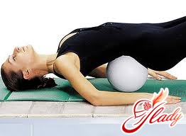
Measures and means of prevention of osteochondrosis
It is also very important to know how you canпредупредить возникновения остеохондроза поясничного отдела. Профилактика заболевания поможет избежать много неприятных минут, связанных с наличием заболевания, его диагностикой и последующим лечением. И, конечно же, нельзя забывать о том. Что профилактика значительно дешевле лечения уже развившегося заболевания. Правильно подобранный рацион питания Питание крайне важно для нормального функционирования всех систем человеческого организма. Не стал исключением и поясничный остеохондроз. Специальная диета не только является замечательным профилактическим средством, но и помогает облегчить состояние уже имеющегося у человека заболевания, тем самым повышая эффективность лечения. Главное условие правильно составленного рациона питания для человека, страдающего любым заболеванием позвоночника, в том числе и остеохондроза поясничного отдела позвоночника, это бессолевое питание. В меню больного человека должны входить такие продукты, как овощи, фрукты, нежирные сорта мяса. Крайне важно полностью исключить из рациона все жирные, острые и жареные продукты, специи, соль и сахар. Из напитков стоит отдать предпочтение чаю, отвару шиповника и брусники. Полностью исключите употребление кофе, газированных и алкогольных напитков. Правильное положение лежа и выбор постели Для профилактики возникновения заболевания и успешного лечения очень важно знать, каким же образом правильно лежать и, главное, на чем. Оптимальным выбором станет ровная и в меру жесткая постель. Не стоит впадать в фанатизм и пытаться спать на досках. Гораздо разумнее покрыть постель тонким щитом, изготовленным из деревянных досок, поверх которого необходимо постелить тонкий матрас. В том случае, если подходящего матраса под рукой не окажется, вы вместо них можете использовать несколько тонких шерстяных одеял. Эта мера необходима для того, чтобы спина восстанавливала свою физиологическую форму, а имеющие место подвывихи позвонков выпрямлялись. Однако будьте готовы к тому, что в первое время вы будете испытывать довольно интенсивные болевые ощущения, которые будут продолжаться до тех пор, пока позвонки не примут нормальное положение. Для облегчения этого состояния первое время можно подкладывать под болезненный сустав валик из ваты. Тем самым вы снимите напряжение мышц и слегка ослабите болезненные ощущения. Очень многие люди, страдающие от остеохондроза поясничного отдела, совершают одну и ту же ошибку – ложатся спать на спину. Однако в данном случае гораздо разумнее будет лечь спать на живот, подтянув под грудь согнутую в коленке ногу. Под живот можно подложить плоскую тонкую подушку. И только пролежав в таком положении хотя бы полчаса, можно очень аккуратно повернуться на спину, завести руки за голову, полностью вытяните ноги и разведите их носками в разные стороны. В том случае, если болевые ощущения слишком сильные и проделать все вышеописанные действия не удается, выполняйте их ровно настолько, насколько вы в состоянии. С каждым разом у вас будет получаться все лучше и лучше. Утром, после пробуждения, вставать с постели человеку, страдающему от остеохондроза, крайне сложно и зачастую больно. Для того чтобы облегчить данный процесс, врачи рекомендуют следующее. После того, как вы проснулись, повернитесь на спину, потянитесь и руками и ногами несколько раз. После этого начните очень аккуратно двигать ступнями ног по часовой стрелке. После этого осторожно повернитесь на живот, еще раз потянитесь и очень аккуратно опустите поочередно ноги на пол. После этого переносите вес на ноги, опираясь на руки. Вставайте также очень аккуратно, не делая резких движений. Не менее важно правильно сидеть Ведь в том случае, если человек сидит неправильно, тяжесть распределяется неравномерно и оказывает крайне негативное влияние на позвоночный столб. Для того чтобы этого не происходило, тело человека должно опираться не на поясницу или копчик, а на седалищные бугры, которые, собственно, для этого и предназначены. Однако подобное возможно только в одном случае – если человек сидит на твердой поверхности. Также очень важно правильно подобрать высоту стула – она должна соответствовать длине голени. Неправильное сидении также входит в основные причины обострения остеохондроза. В том случае, если по работе вы вынуждены проводить сидя длительное количество времени, каждые полчаса совершайте повороты корпуса в обе стороны. Также обязательно совершайте по пять круговых вращений, как шеей, так и плечами. Следите за тем, чтобы плечи были максимально развернуты, а голову старайтесь держать максимально прямо. Не менее пристального внимания заслуживает сидение за рулем. Спина должна иметь полноценную опору. Купите специальный валик, который необходимо постоянно прокладывать между спинкой сидения и поясницей. Во время езды держите спину и голову прямо. Не проводите за рулем более 3 часов подряд. Обязательно совершайте регулярные остановки. Выходите из машины и совершайте простые физические упражнения, такие как поднимание и опускание рук, приседания, повороты и наклоны. В конце концов, даже простое хождение вокруг машины способно оказать положительное воздействие на состояние позвоночника и мышечной системы. То же самое касается и просмотра телевизора либо чтения. Самое главное правило – не задерживайтесь подолгу в одной и той же статической позе – это крайне негативно влияет на состояние позвоночника. Многие люди пытаются использовать народные методы лечения остеохондроза. Однако этого делать все же не стоит, так как позвоночник – слишком серьезное и сложное явление. И ни в коем случае не стоит ставить эксперименты, гадая, поможет ли тот или иной рецепт народной медицины. Ведь в случае неудачи цена ошибки будет слишком высокой. В лучшем случае улучшения просто не наступит. А в худшем – человек может поплатиться за ошибку возможностью ходить. В том случае, если вы или ваши близкие заметили наличие каких – либо проблем со стороны позвоночника, вам необходимо как можно быстрее обратиться за помощью к врачам, а не заниматься самолечением. Возможности современной медицины достаточно широки, и излечению поддается практически любое заболевание. Главное, вовремя его диагностировать и начать лечение. Именно поэтому ни в коем случае нельзя затягивать визит к врачу. Остеохондроз, как и любое другое заболевание, наиболее успешно лечится именно в самом начале заболевания, пока к нему не присоединились другие осложнения, типичные для данного заболевания. В таком случае лечение остеохондроза значительно усложняется и занимает гораздо больше времени. Советуем почитать:


